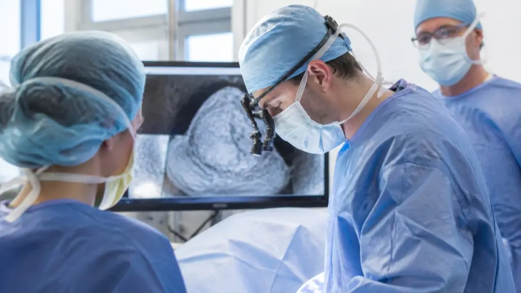
Mohs skin cancer surgery uses a stepwise approach to remove skin cancer. The surgeon removes a thin layer of tissue, and they examine it to see whether cancer cells remain. If residual cancer appears, the surgeon returns only to the precise area where those cells were found, instead of taking a broad, deep section. Here is some information about how imaging techniques support Mohs surgery:
Maps Cancer Margins
Advanced imaging helps map cancer margins before and during Mohs skin cancer surgery. This mapping supports decisions about how far to extend each stage. High-resolution dermoscopy, confocal microscopy, and other imaging tools give detailed views of tumor borders. The surgeon then correlates those images with surface markings on the skin. When imaging highlights subtle extensions of the lesion beyond what the eye sees, the surgical plan adapts, and the surgeon can target more of the tumor. This allows the surgeon to remove affected tissue more precisely while preserving as much healthy skin as possible.
Reduces Surgical Stages
When the preoperative map is more accurate, Mohs surgery sometimes requires fewer stages. This often shortens treatment times. Imaging supports a more targeted first pass by providing a clearer picture of the lesion’s depth and spread. That information shapes the initial margins, and it guides the surgeon toward areas that deserve closer attention during microscopic review. Imaging may also help reduce additional stages by:
- Helping surgeons recognize complex tumor patterns that make a procedure more complicated
- Providing reference images that can be compared with findings from each stage
- Helping the surgical team anticipate areas where tumor growth is difficult to assess
- Supporting more informed decisions about whether another stage is necessary
Preserves Healthy Tissue
By visualizing structures beneath the surface, imaging helps distinguish tumor tissue from nearby normal skin. This matters in areas such as the nose, eyelids, lips, and ears. When the surgeon has a clearer view of where the cancer stops, each stage removes only the tissue that corresponds to mapped tumor spread. This selective approach protects healthy collagen and vessels, and it gives reconstruction a stronger foundation.
Digital imaging also supports reconstruction planning because it documents the size and orientation of the defect once all tumor tissue is cleared. The surgeon reviews these images while choosing between options such as linear closure, local flaps, or skin grafts. This planning often matches the natural lines and contours of the face. Imaging records serve as a reference during follow-up visits, and they help track how the scar matures over time.
Schedule Mohs Skin Cancer Surgery
Imaging techniques provide surgeons with additional information that supports more precise Mohs surgery. By improving the visibility of tumor margins, these tools guide surgical stages and limit the removal of healthy tissue. They also give surgeons details for planning reconstruction and monitoring healing after the procedure. Although imaging does not replace the microscopic examination of tissue, it can strengthen decisions throughout the process. Contact a dermatologist today to schedule a skin exam and learn more about options for skin cancer surgery.